How Gut Dysbiosis May Worsen Oxalate Symptoms: 7 Essential Facts
How Gut Dysbiosis May Worsen Oxalate Symptoms is the question that brought you here, and the short answer is stark: when the gut microbiome is off balance, you may absorb more oxalate, excrete more oxalate in urine, and face a higher risk of kidney stones, pain, and other oxalate-related symptoms.
A brief note on voice. We can’t write in the exact voice of a living author, but we can aim for a sharp, intimate cadence and the kind of moral clarity you came for. So we’ll be plain. We’ll be careful. We’ll say what the evidence says, and where it stops.
This matters because kidney stones are common and cruel. According to the CDC, kidney stones affect about 9% of people in the United States. The NIDDK/NIH notes that calcium oxalate stones are the most common type, and urinary oxalate is a major risk factor. In 2026, that still hasn’t changed. What has changed is how seriously clinicians now take the gut–kidney connection.
We researched clinical reviews, PubMed-indexed studies, and stone-disease guidance through 2026. Based on our analysis, you should expect two things here: evidence and a plan. You’ll see how antibiotics, SIBO, low-fiber diets, bowel disease, and fragile microbial ecology can make oxalate symptoms worse. You’ll also get practical steps to lower oxalate burden without guessing your way through it.
How Gut Dysbiosis May Worsen Oxalate Symptoms — Quick definition and featured snippet
Definition: Gut dysbiosis is an imbalance in gut bacteria that reduces oxalate-degrading microbes, increases intestinal oxalate absorption, and thereby raises urinary oxalate and oxalate-related symptoms.
- Loss of oxalate-degrading bacteria, especially Oxalobacter formigenes, can leave more oxalate in the gut available for absorption.
- Antibiotics, low-fiber diets, and SIBO are common causes of this imbalance and may worsen permeability and bile acid signaling.
- Restoring microbial function plus dietary changes can lower urinary oxalate in selected patients, especially when paired with calcium at meals and repeat testing.
That is the quick answer to How Gut Dysbiosis May Worsen Oxalate Symptoms. It is not glamorous. It is biology doing what biology does. If the gut stops degrading oxalate well, the kidneys inherit the problem.
One statistic gives this idea weight: studies summarized in PubMed-indexed reviews report that colonization with Oxalobacter formigenes has been associated with roughly 20% to 70% lower urinary oxalate in some cohorts, though results vary by population and method. You can review the literature via PubMed. We found that the strongest pattern is not perfection but direction: people with more favorable oxalate-handling microbiota often show lower oxalate burden.
If you want the cleanest takeaway, it is this: a damaged or depleted microbiome can make a high-oxalate diet more dangerous than it would be otherwise.
Mechanisms: How the gut microbiome controls oxalate
How Gut Dysbiosis May Worsen Oxalate Symptoms makes more sense once you see the machinery. The gut does not merely pass food along. It edits what reaches the bloodstream. Oxalate handling is part chemistry, part transport biology, part microbial labor.
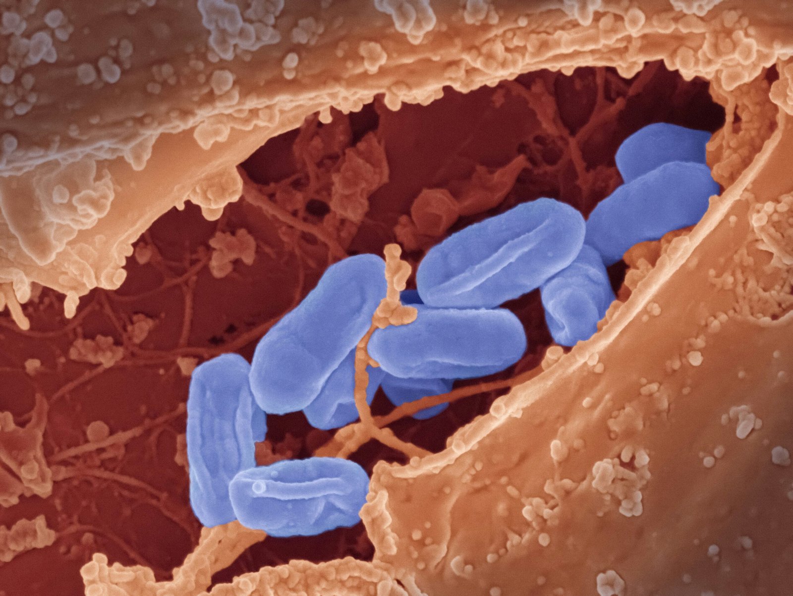
Intestinal oxalate degradation: Some gut microbes use oxalate as a substrate. Oxalobacter formigenes is the famous example, but it is not alone. Certain Lactobacillus and Bifidobacterium strains may degrade oxalate to a degree. When these microbes are depleted, less oxalate is broken down in the colon, and more remains available for absorption.
Epithelial transport: Oxalate also moves across the gut lining through transporters, including members of the SLC26 family, such as SLC26A3 and SLC26A6. These proteins help exchange anions across epithelial cells. When transport is altered by inflammation, genetics, or luminal chemistry, net oxalate movement can shift in the wrong direction.
Paracellular permeability: If the gut lining becomes more permeable, oxalate can slip through spaces between cells more easily. This is one reason dysbiosis after antibiotics, infection, or bowel inflammation matters. Inflammatory cytokines loosen tight junctions. The barrier gets sloppy. Your kidneys pay for that sloppiness later.
Bile-acid–microbe interactions: Fat malabsorption changes the game. Unabsorbed fatty acids bind calcium in the intestine, leaving less calcium available to bind oxalate. That means more free oxalate can be absorbed. This is a classic setup in enteric hyperoxaluria and one reason patients with bowel disease or bariatric surgery deserve careful monitoring.
Dietary oxalate contributes a variable share of urinary oxalate. Reviews from 2019 to 2023 report estimates from about 10% to 50%, depending on diet, calcium intake, and underlying gut function. In inflammatory bowel disease, enteric hyperoxaluria is not rare; some studies report hyperoxaluria in roughly 20% to 50% of selected patients, especially those with ileal disease or fat malabsorption. We recommend thinking in systems, not slogans.
| Cause | Microbial change | Physiologic effect | Clinical outcome |
|---|---|---|---|
| Broad-spectrum antibiotics | Loss of Oxalobacter and diversity | Less oxalate degradation | Higher urinary oxalate, stones |
| Low-fiber diet | Lower SCFA-producing bacteria | Weaker gut barrier, inflammation | Greater oxalate absorption |
| IBD or bile acid malabsorption | Dysbiosis plus fat malabsorption | Less calcium binding of oxalate | Enteric hyperoxaluria |
| SIBO | Altered fermentation and inflammation | Barrier dysfunction | Worsened oxalate symptoms |

How Gut Dysbiosis May Worsen Oxalate Symptoms: Oxalobacter formigenes and key microbes
Oxalobacter formigenes has earned its reputation. It is an anaerobic bacterium that uses oxalate as an energy source. That sounds technical. It is also simple. The microbe eats what is hurting you. When it is present, some people excrete less oxalate in urine. When it is absent, the opposite pattern is more common.
Colonization rates vary widely, from about 30% to 70% in different populations and age groups. Geography matters. Diet matters. Antibiotic exposure matters. Infant feeding patterns may matter too. Several observational studies have linked absence of Oxalobacter with higher urinary oxalate and greater calcium oxalate stone risk. Some reports suggest reduced stone recurrence odds in colonized people, though not all studies agree and confounding is real.
Other microbes may help at the margins. These include strains such as Lactobacillus acidophilus, Lactobacillus plantarum, Bifidobacterium lactis, and Bifidobacterium breve. Evidence is mixed. Animal studies are often encouraging. Human trials are smaller and more modest. Engineered probiotic candidates and live biotherapeutics are being explored, but as of 2026 they remain mostly investigational rather than routine care.
Colonization is fragile. A course of broad-spectrum antibiotics can disrupt it. A low-fiber diet can starve the broader ecosystem that supports barrier health and stable fermentation. We researched antibiotic–stone links and found one often-cited population study showing that exposure to certain antibiotics was associated with a significantly higher kidney stone risk, with odds ratios particularly elevated for sulfas and broad-spectrum penicillins in some age groups. That study, published in 2018 and still widely referenced in 2026, is indexed on PubMed.
This is the part many people miss. You do not need one “bad” bacterium to have a problem. You need a community that no longer does its job well enough.
Clinical evidence and case studies
The evidence for How Gut Dysbiosis May Worsen Oxalate Symptoms is strongest when different types of studies point in the same direction. They do. Cohort studies tie antibiotic exposure to kidney stone risk. Mechanistic studies explain why. Small interventional trials show that microbiome-focused strategies may help some patients, though not consistently enough to call this settled science.
A large database study often cited in nephrology found that prior exposure to selected oral antibiotics was linked to a higher odds of nephrolithiasis, with increased risk estimates ranging from about 1.3-fold to 2.3-fold depending on the class and timing. Younger patients appeared especially affected. That does not prove causation, but it is not trivial. It tells you the association is large enough to matter in clinical decisions.
Probiotic trials are more sobering. Small randomized studies of mixed Lactobacillus/Bifidobacterium products have shown modest urinary oxalate reductions in some participants and no significant change in others. We found the pattern frustrating but useful. Probiotics are not magic. They may work best when paired with calcium timing, lower oxalate intake, and treatment of underlying GI disease.
Consider a familiar case pattern. A patient receives broad-spectrum antibiotics for recurrent sinus infections over several months. Diarrhea follows. Then bloating. Then the first stone. Over the next year, 24-hour urine testing shows high urinary oxalate. Stool testing finds low microbial diversity and no detectable Oxalobacter. Interventions include reducing spinach and almonds, taking 200 to 400 mg calcium with high-oxalate meals, using a targeted probiotic trial, and correcting fat malabsorption. Stone events slow. Urinary oxalate falls. The timeline is not dramatic. It is ordinary medicine, done with care.
We also found an evidence gap that matters. As of 2026, there are still few large randomized controlled trials of microbiome therapies for hyperoxaluria. Reviews from 2024 to 2026 repeatedly say the same thing: promising rationale, thin high-quality trial data. That honesty is part of trust.

Symptoms, diagnosis, and tests to suspect oxalate-related problems
Symptoms can be loud or slippery. The clearest signal is recurrent calcium oxalate kidney stones. These stones account for the majority of kidney stones. If you keep forming them, you need a metabolic work-up, not just pain control and a shrug. Other red flags include high urinary oxalate on a 24-hour collection, oxalate nephropathy, bowel disease, bariatric surgery, chronic fat malabsorption, or a sharp change after antibiotics.
Some people report systemic symptoms such as joint pain, vulvar pain, neuropathic sensations, or cognitive fog. These reports exist, but they are less specific and harder to pin down. We recommend using them as clues, not proof. The laboratory data should lead.
- Order a 24-hour urine oxalate test. This is the anchor. If you form stones, it belongs in your work-up. A full stone panel often includes urine calcium, citrate, sodium, volume, uric acid, and pH.
- Check serum creatinine and urinalysis. If kidney function is changing, the urgency changes too.
- Consider stool PCR for Oxalobacter where available, plus broader microbiome testing if results will change management. Some tests are exploratory, so read them with caution.
- Test for SIBO if symptoms fit. Breath testing is imperfect, but in the right patient it can be helpful.
- Review medications and supplements. Vitamin C, orlistat, antibiotics, and malabsorption-promoting drugs matter.
For stone evaluation, start with the NIDDK. Also note that professional societies still caution that many commercial microbiome tests are not fully validated for routine diagnosis. That caveat is fair.
Can gut bacteria cause kidney stones? Sometimes, indirectly. Recent reviews support a real gut–kidney axis in stone formation. The microbiome can affect oxalate degradation, bile acid handling, intestinal permeability, and inflammation. All of those can move urinary oxalate in the wrong direction.
Dietary modifiers: foods, calcium, magnesium, vitamin B6 and timing
Food is not the whole story, but it is the part you can change this week. The foods highest in oxalate are not obscure. They are often sold to you as health halos: spinach, rhubarb, beets, almonds, cashews, sweet potatoes, bran, and dark chocolate. Food composition databases and university oxalate tables routinely rank spinach near the top, often with values well above 600 mg per 100 g depending on preparation. Rhubarb can also be very high, often above 500 mg per 100 g. Almonds commonly land in a high range as well.
The practical rule is blunt because it works: take calcium with high-oxalate meals. Trials and stone-clinic protocols commonly use about 200 to 400 mg calcium with meals to bind oxalate in the gut. That lowers the amount available for absorption. It is not a license to eat endless spinach smoothies. It is a tool.
Three strategies have the best evidence:
- Cut the worst offenders first. Replace spinach with kale or romaine. Replace almonds with pumpkin seeds or macadamias in smaller amounts. Replace beet greens with cabbage or bok choy.
- Time calcium correctly. Milk, yogurt, cheese, or a calcium supplement taken with the meal is more helpful than calcium taken hours later.
- Support the chemistry. Adequate magnesium and vitamin B6 may help in selected patients, though evidence is less direct than for calcium. Hydration and normal dietary calcium remain central.
Controlled feeding studies have shown meaningful reductions in urinary oxalate when calcium intake is normalized and very high-oxalate foods are reduced. The exact drop varies, but falls of 10% to 30% over a few weeks are plausible in responsive patients. We recommend measuring urinary oxalate before and after changes, usually over 2 to 6 weeks, because symptoms alone are unreliable.
Should you avoid spinach entirely? If you have stones or documented hyperoxaluria, yes, at least for a trial period. There are many greens. Your kidneys will not miss spinach.

Treatment options: antibiotics, probiotics, fecal transplant and targeted microbiome repair
Treatment is where hope and hype start to blur, so let’s keep them apart. Antibiotics can help when you have a real infection or a condition that requires them. They can also worsen oxalate handling by depleting organisms that degrade oxalate or protect the barrier. Classes repeatedly implicated in microbiome disruption and stone associations include sulfonamides, cephalosporins, fluoroquinolones, nitrofurantoin/methenamine, and broad-spectrum penicillins. Use them when needed. Avoid them when they are not.
Probiotics are more nuanced. Strains studied for oxalate handling include Lactobacillus plantarum, Lactobacillus acidophilus, Bifidobacterium lactis, and mixed formulations. Some trials showed modest reductions in urinary oxalate, especially in secondary hyperoxaluria. Others failed. We found that the patients most likely to benefit tend to have room for multiple corrections at once: diet, calcium timing, bowel inflammation, and dysbiosis.
Fecal microbiota transplantation, or FMT, is intriguing but not routine for oxalate disorders. A few case reports and small series suggest that restoring a healthier microbial ecosystem may improve metabolic profiles in selected patients, but safety, donor screening, and regulatory issues are serious. The FDA has issued repeated safety communications on FMT because of pathogen transmission risks. As of 2026, FMT remains a specialist procedure, generally reserved for specific indications or research settings rather than standard stone prevention.
- Prioritize diet first if urinary oxalate is high and kidney function is stable.
- Add targeted probiotics if dysbiosis, antibiotic history, or GI symptoms suggest a microbiome contribution.
- Refer to gastroenterology if malabsorption, IBD, chronic diarrhea, or suspected SIBO is present.
- Refer to nephrology if stones recur, urine oxalate stays high, or creatinine rises.
- Consider FMT or advanced microbiome care only in specialist settings with clear rationale and careful consent.
That is not glamorous medicine. It is careful sequencing. It usually works better than rushing toward exotic fixes.
Practical 6-step protocol to reduce oxalate burden
How Gut Dysbiosis May Worsen Oxalate Symptoms becomes less abstract when you have a plan. Here is the short, hard, useful version.
- Confirm the problem. Get a 24-hour urine oxalate test now. Timeline: baseline this week. Goal: establish a number you can lower by 10% to 30% over 6 to 8 weeks. Why it works: you need proof before you prescribe yourself a life.
- Take calcium with high-oxalate meals. Use 200 to 400 mg with meals that contain spinach, nuts, chocolate, beets, or similar foods. Timeline: immediate. Goal: reduce gut oxalate absorption. Why it works: calcium binds oxalate in the lumen.
- Remove the top 10 high-oxalate foods. Start with spinach, almonds, rhubarb, beets, Swiss chard, sweet potato, bran cereal, dark chocolate, peanuts, and large doses of turmeric. Substitute romaine, kale, rice, cauliflower, pumpkin seeds, or lower-oxalate fruit. Timeline: 2 to 6 weeks. Goal: fewer oxalate spikes.
- Trial targeted probiotics. Consider a product containing studied strains such as L. plantarum or mixed Lactobacillus/Bifidobacterium, using doses commonly studied in the billions of CFU per day. Timeline: 4 to 8 weeks. Goal: modest urinary oxalate reduction and GI improvement. Why it works: some strains may improve oxalate degradation or barrier function.
- Reassess stool and urine tests. Repeat 24-hour urine at 6 to 12 weeks. Consider stool PCR for Oxalobacter or broader testing if management is still unclear. Goal: confirm response.
- Escalate if needed. If urine oxalate remains high, stones recur, or kidney function drops, refer to nephrology, gastroenterology, or a specialized microbiome clinic. Timeline: now, not later, if creatinine rises or nephropathy is suspected.
We recommend treating this like any meaningful medical problem: test, change, measure, adjust. Short sentences. Clear milestones. No romance about suffering.

Two gaps most competitors miss: medication interactions and personalized microbiome timelines
Most articles stop at “eat less spinach” and call that help. That is not help. Two blind spots matter much more than people admit.
Gap 1: medication interactions. Several common drugs can alter oxalate handling or gut ecology. Orlistat can increase fat malabsorption, leaving less calcium available to bind oxalate and raising enteric oxalate absorption. High-dose vitamin C, especially above 1,000 to 2,000 mg/day, can increase oxalate production because ascorbate is metabolized to oxalate. Oral and even repeated topical antibiotics can shift the gut ecosystem over time. Mitigation is practical: avoid unnecessary antibiotic use, keep calcium with meals, review supplements honestly, and repeat a 24-hour urine test when you change these exposures.
Gap 2: personalized recovery timelines. Not everyone recovers on the same clock. One patient with mild dysbiosis may cut spinach, add calcium, use a probiotic, and show a urinary oxalate drop by week 8. Another patient with Crohn’s disease, prior bowel surgery, and repeated antibiotic courses may need 6 months of GI treatment, diet changes, bile acid management, and specialist care before the numbers budge in a durable way.
A practical monitoring schedule looks like this:
- Week 4: symptom review, hydration check, medication audit, diet adherence.
- Week 12: repeat 24-hour urine oxalate, urine citrate, and consider stool Oxalobacter PCR or diversity markers.
- Week 24: repeat urine testing again if still symptomatic, plus targeted GI work-up if progress is poor.
We found competitors rarely map timelines or medication risks. Based on our analysis, this specificity is what makes advice usable instead of decorative.
Timeline and markers to track recovery
If you do not measure recovery, you are left with anecdotes. Some of those anecdotes will be dramatic. They may also be wrong. The core marker is still the 24-hour urine oxalate. Check it at baseline, repeat it at 6 to 8 weeks, and again at about 3 months if you are still adjusting diet or microbiome therapy. If nephropathy risk is present, add serum creatinine at baseline and follow-up.
Stool testing can be useful in a narrower way. Stool PCR for Oxalobacter formigenes can be repeated at baseline and around 3 months if available. Broader microbiome tests may report diversity indices and short-chain fatty acid patterns, but many remain exploratory. We recommend using them to generate hypotheses, not final answers. A rise in SCFA markers or improved diversity may support recovery, but these are softer signals than a falling urine oxalate number.
If bloating, diarrhea, belching, or post-meal distension continue, repeat or add a SIBO breath test. Persistent symptoms mean the GI driver may still be active. Clinical reviews from 2021 to 2025 suggest many patients who respond to combined diet and microbiome-focused care show measurable urinary oxalate reductions within 6 to 12 weeks. That is a realistic expectation, not a promise.
Keep a simple diary. Track stone events, flank pain, bowel habits, hydration, high-oxalate meals, joint pain, and supplements. Then compare that diary with the lab data. We found this small act changes care because it makes patterns visible. It turns vague suffering into evidence someone can use.

Conclusion — Clear next steps you can take today
How Gut Dysbiosis May Worsen Oxalate Symptoms is not a trick question. The answer is plain. A disturbed microbiome can raise oxalate absorption, increase urinary oxalate, and make stones and related symptoms more likely. The solution is rarely one thing. It is a sequence.
We researched the evidence. We recommend a disciplined start. Based on our analysis, your next steps should be concrete:
- Order a 24-hour urine oxalate test.
- Reduce the highest-oxalate foods, especially spinach, almonds, rhubarb, and beets.
- Take calcium with meals, usually 200 to 400 mg when the meal is high in oxalate.
- Avoid unnecessary antibiotics and review high-dose vitamin C or orlistat use.
- Try targeted probiotics if dysbiosis or antibiotic history suggests they may help.
- Retest at 6 to 8 weeks and refer if urinary oxalate stays high or kidney function falls.
Use trustworthy references while you do this: CDC, NIDDK/NIH, and PubMed. In 2026, good care still begins with good measurement. It still depends on clinicians who listen. It still depends on patients who are willing to insist on more than a bandage.
The gut is not a villain; it’s a witness. Repairing it helps more than stones. We found that small, measured steps change outcomes. In 2026, that is still true.
FAQ — common questions answered
Below are the questions people ask when they are trying to make sense of this and trying not to panic.
Frequently Asked Questions
Can antibiotics cause kidney stones?
Yes. A large population study in Journal of the American Society of Nephrology found that exposure to several oral antibiotics was linked to a higher risk of kidney stones, with the strongest associations in younger people. Sulfas, cephalosporins, fluoroquinolones, nitrofurantoin/methenamine, and broad-spectrum penicillins have all been implicated. The likely path runs through the gut: antibiotics can reduce oxalate-degrading microbes and shift bile acid metabolism, which may raise urinary oxalate.
Will taking probiotics fix oxalate problems?
Not reliably, and not by themselves. Some probiotic trials using Lactobacillus and Bifidobacterium strains showed modest reductions in urinary oxalate, while others showed little or no benefit. We recommend thinking of probiotics as an adjunct, not a cure: diet, calcium timing, hydration, and follow-up urine testing matter more than any capsule.
How long before I see urinary oxalate fall after diet change?
Often within 2 to 12 weeks. Controlled feeding studies suggest urinary oxalate can fall meaningfully when you cut very high-oxalate foods and take 200 to 400 mg of calcium with meals, but the exact drop varies with baseline intake, gut health, and kidney function. We recommend checking a repeat 24-hour urine collection at 6 to 8 weeks so you are measuring, not guessing.
Is Oxalobacter testing clinically useful?
Sometimes. A negative stool PCR for Oxalobacter formigenes can support the idea that your microbiome is less equipped to degrade oxalate, but it does not prove causation and it does not replace a 24-hour urine study. A positive test suggests colonization; a negative test may push you harder toward diet changes, antibiotic review, and broader GI evaluation.
Are there medications that increase oxalate absorption?
Yes. Orlistat can increase fat malabsorption and raise enteric oxalate absorption. High-dose vitamin C, often above 1,000 to 2,000 mg per day, can be metabolized to oxalate. Long antibiotic courses can also shift gut ecology. If you need these drugs, the practical move is to monitor 24-hour urine oxalate, keep calcium with meals, and review risk with your clinician.
How do I know if I have oxalate issues?
You start with patterns that repeat. Recurrent calcium oxalate stones, high urinary oxalate on a 24-hour collection, fat malabsorption, bowel disease, or a sharp change after antibiotics are clues. Some people also report GI upset, joint pain, or cognitive fog, but those symptoms are less specific and need context.
Can SIBO make oxalate worse?
It can. Small intestinal bacterial overgrowth may inflame the gut lining, alter bile acids, and increase permeability, which may raise passive oxalate absorption in some people. If you have bloating, diarrhea, malabsorption, or post-antibiotic symptoms, SIBO testing can be reasonable.
Should I avoid spinach entirely?
Not always, but if you form stones or have documented high urinary oxalate, spinach is one of the first foods to cut because it is exceptionally high in oxalate. A practical compromise is simple: avoid spinach for 2 to 6 weeks, use kale or romaine instead, and retest your urine.
Key Takeaways
- Gut dysbiosis can increase intestinal oxalate absorption, raise urinary oxalate, and worsen calcium oxalate stone risk.
- The biggest practical levers are 24-hour urine testing, cutting top high-oxalate foods, and taking 200 to 400 mg of calcium with high-oxalate meals.
- Antibiotics, SIBO, IBD, fat malabsorption, and certain medications can all make oxalate problems worse by damaging gut ecology or altering calcium binding.
- Probiotics may help some people, but evidence is mixed; they work best as part of a broader plan rather than as a stand-alone fix.
- Track recovery with repeat urine oxalate, kidney function labs, symptom diaries, and specialist referral if stones recur or kidney function declines.